Home ![]() MAYFAIR DIAGNOSTICS' VASCULAR LAB IS MOVING
MAYFAIR DIAGNOSTICS' VASCULAR LAB IS MOVING
For almost 20 years, the Mayfair Diagnostics Vascular Lab at Mayfair Place has been offering sub-specialized, multidisciplinary diagnostic imaging that is non-invasive to care for our patients’ vascular needs. In mid-October 2021, the Vascular Lab will be moving from Mayfair Place to our newest clinic location – Mayfair Diagnostics Sunridge.
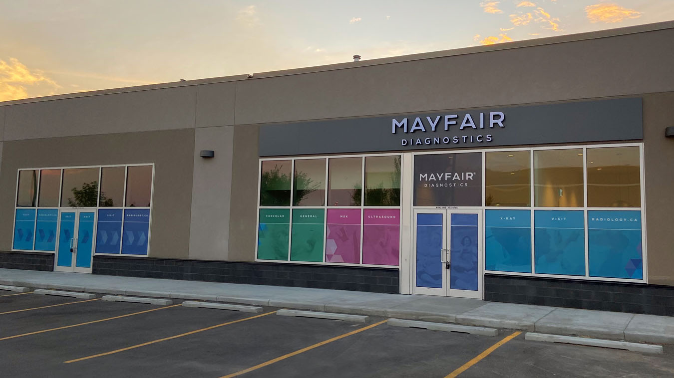
Located in unit 150 at 3363 – 26th Avenue NE, our new Sunridge clinic will be across from the Peter Lougheed Centre, beside the Best Buy, and between Dental Care for Children and the Five Guys restaurant. Mayfair Diagnostics will be making this move to better serve our patients. It’s especially ideal for vascular surgery patients who have same-day appointments at the Peter Lougheed Centre.
Effective September 8, patients will be able to call our Contact Centre (403-777-3000) to book ultrasound exams at our Sunridge clinic for October. Walk-in X-ray services will not be available until October when the clinic is officially open.
Please note that every vascular referral is triaged by our vascular team to ensure the most appropriate testing is performed for optimal patient care. Our experienced vascular team includes sub-specialized vascular interventional radiologists, registered vascular technologists, and an accredited clinical vascular scientist with an MSc in vascular imaging who will also be the Sunridge clinic manager. This team ensures that our patients receive expert imaging that is appropriate for their medical history, and quality reports that help ensure an accurate diagnosis.
Using sound waves, a vascular ultrasound exam looks at the blood flow in your veins and arteries. High-frequency sound waves are transmitted from a handheld device, called a probe or transducer, which is placed on the body and moved around the area of interest. A gel is applied to the skin to aid in the movement of the transducer. The transducer collects the sounds that bounce back and a computer then uses those sound waves to create an image on a computer screen. Ultrasound exams are noninvasive, don’t use radiation, and images are captured in real-time, showing the movement of your blood flowing through your blood vessels.
At many of our clinics we perform two common vascular studies:
At our Vascular Lab, our experienced and highly collaborative team can examine complex medical histories and provide the best results for our patients. Here are examples of exams available at our Vascular Lab, which will continue to be offered at our Sunridge location:
For some vascular ultrasound exams, you will be asked to lie on your back and blood pressure cuffs will be place on both arms and both ankles. The blood pressure cuffs will inflate so the sonographer can take blood pressure readings. The cuffs may become tight, but only for a few seconds. Other exams don’t require cuffs, the transducer will simply be passed over the area of concern.
During the exam your sonographer will listen to your blood flow, so you may hear what sounds like your heartbeat. This is the blood moving through the blood vessel and each blood vessel checked has a slightly different sound. A warm, non-scented, hypo-allergenic ultrasound gel will be applied to the area being scanned and a series of images will be acquired to show the structure and flow of blood through the blood vessels. Depending on the type of exam, you may be asked to move your arms, lie on your side, sit, or stand to help acquire the best images of the area in question.
Exams can range from 30 minutes to one hour depending on their complexity. Please visit the vascular ultrasound services page for information on specific exams.
After your exam a specialized radiologist will review your images and interpret them before forwarding a report to your referring physician. If there are any concerns with your results, a radiologist may speak with you or fax a report to your doctor. For non-urgent results, your doctor will receive a detailed report, which will outline your diagnosis, by the next business day following your exam. Your doctor will then discuss your results with you.
Ultrasound is non-invasive and safe. Occasionally, an ultrasound exam may require pressure to ensure the best image of the area in question and may be temporarily uncomfortable, such as when an area of tenderness is scanned. As well, during a venous ultrasound the transducer will be pressed against your leg to cause the veins to squeeze closed momentarily. This is not harmful to the vein, but it’s the preferred method of checking for a blood clot. The pressure may be quite uncomfortable for the few seconds it takes to evaluate each segment of the vein down the leg.
Vascular ultrasound imaging must be requested by a health care practitioner who will provide a requisition. Your medical and family history, risk factors, and type and duration of symptoms, all affect a referring physician’s decision on which type of imaging is appropriate.
When we receive your requisition Mayfair Diagnostics will contact you to schedule your exam and provide you with detailed information to prepare for it. Once your exam is completed, your images will be reviewed by a specialized radiologist who will compile a report that is sent to your doctor.
Venous and carotid artery ultrasounds are performed at most Mayfair locations in Calgary, while the more complex procedures are performed at our Vascular Lab at our Sunridge location.
© 2022 Mayfair. All rights reserved.
| Cookie | Duration | Description |
|---|---|---|
| cookielawinfo-checkbox-analytics | 11 months | This cookie is set by GDPR Cookie Consent plugin. The cookie is used to store the user consent for the cookies in the category "Analytics". |
| cookielawinfo-checkbox-functional | 11 months | The cookie is set by GDPR cookie consent to record the user consent for the cookies in the category "Functional". |
| cookielawinfo-checkbox-necessary | 11 months | This cookie is set by GDPR Cookie Consent plugin. The cookies is used to store the user consent for the cookies in the category "Necessary". |
| cookielawinfo-checkbox-others | 11 months | This cookie is set by GDPR Cookie Consent plugin. The cookie is used to store the user consent for the cookies in the category "Other. |
| cookielawinfo-checkbox-performance | 11 months | This cookie is set by GDPR Cookie Consent plugin. The cookie is used to store the user consent for the cookies in the category "Performance". |
| viewed_cookie_policy | 11 months | The cookie is set by the GDPR Cookie Consent plugin and is used to store whether or not user has consented to the use of cookies. It does not store any personal data. |