Home ![]() WHY WOULD I NEED DIAGNOSTIC BREAST IMAGING?
WHY WOULD I NEED DIAGNOSTIC BREAST IMAGING?
Diagnostic breast imaging is a type of medical imaging requested to investigate the cause for symptoms in breast tissue, such as lumps, pain, or redness. For both men and women, this often involves a breast ultrasound, a mammogram, or both.
When investigating breast concerns, men under the age of 17 would usually receive a diagnostic breast ultrasound only. For men age 18 and over, your doctor would mostly likely order diagnostic mammography combined with supplemental breast ultrasound imaging.
For women under age 35, investigation of new symptoms would likely involve a diagnostic breast ultrasound only. If you are a woman age 35 and older, a mammogram would be appropriate for new concerns. It might also be combined with supplemental breast ultrasound imaging.
Please note that if you have a family history of breast cancer and are within 10 years of the age a first-degree relative (parent, sibling) was diagnosed with breast cancer, you could potentially be sent for a diagnostic mammogram. For example, if you are 27 and your mother was diagnosed at age 34, a mammogram possibly followed by additional supplemental ultrasound imaging might be requested. Usually, the earliest age this might happen is age 25.
Women age 40 and older may also choose to have regular screening mammography exams – this occurs when there is no obvious breast abnormality and no signs of breast cancer. Having regular screening mammograms makes it easier to compare images and see changes within the breast that might indicate cancer, allowing for early detection.
During a screening mammogram, four images are taken. Occasionally you may be asked to return for additional breast imaging, such as a diagnostic mammogram and/or diagnostic breast ultrasound. This is done to get a more detailed look at any areas of concern and ensure you have received a complete breast exam.
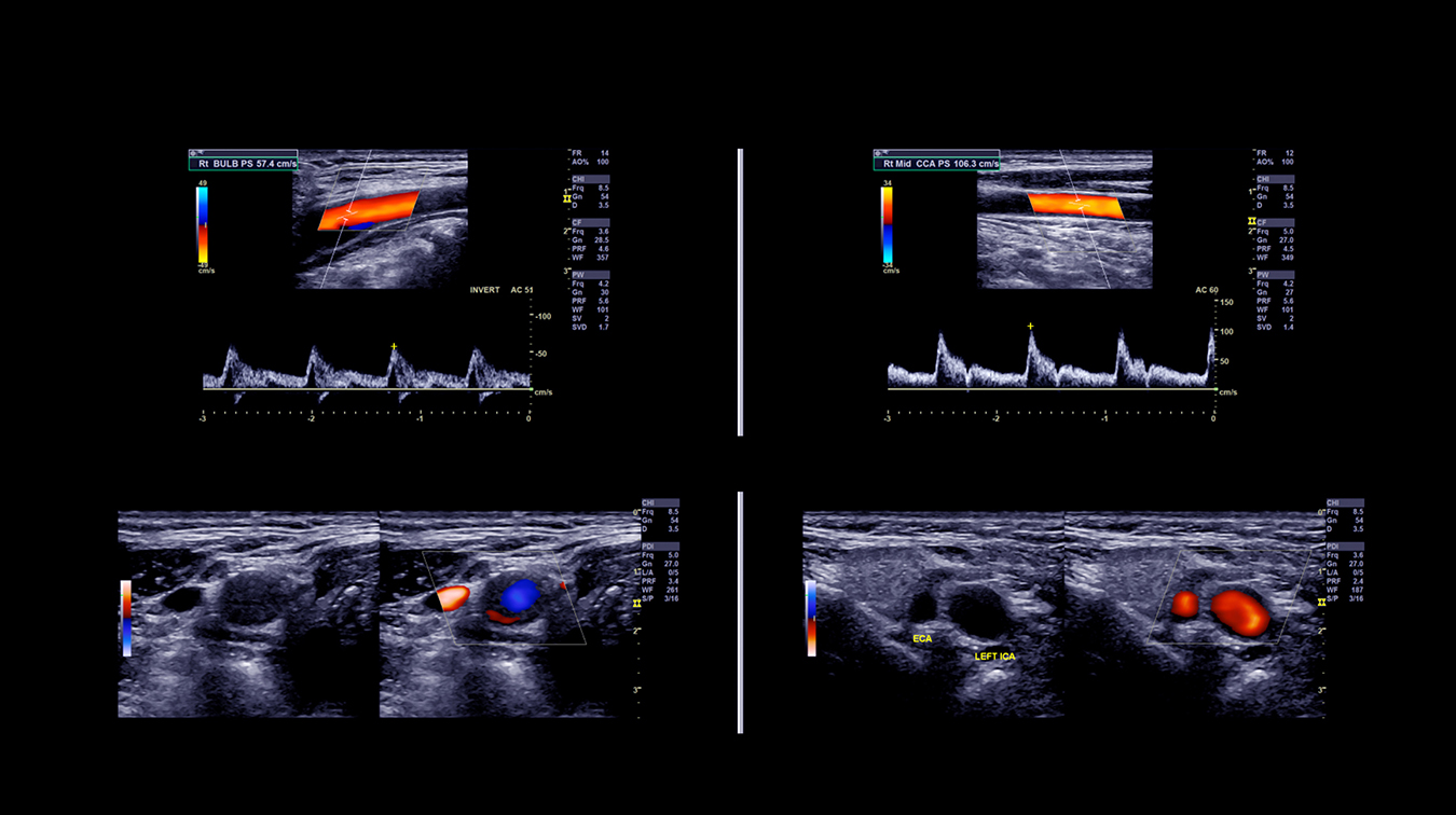
A breast ultrasound can help determine the composition of a lump, distinguishing between a cyst, fibroglandular tissue, and a solid mass. It uses high-frequency sound waves that travel into the breast until they hit a boundary between tissues, such as fluid and soft tissue, or soft tissue and bone. At these boundaries some of the sound waves are reflected back to the probe, while others travel farther until they reach another boundary and are reflected back. Since the pitch, direction, and distance sound waves travel differ depending on the boundary they run into, a computer can interpret this information as a two-dimensional image on a screen.
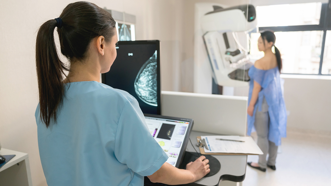
Mammography is a type of X-ray exam that takes an image of the inside of the breasts – called a mammogram. It’s the best way to detect breast cancer in its early, most treatable stage, because it provides a detailed look at the internal structure of breast tissue in both men and women and can reveal changes that are too small to feel.
When used together a mammogram and breast ultrasound can help provide a comprehensive look at breast tissue.
A biopsy can be performed in cases where ultrasound or mammography cannot differentiate benign from malignant lesions. This involves the insertion of a needle into the lesion to take a small tissue sample, which is then sent to a laboratory for analysis.
Factors that can increase the risk of developing breast cancer for both men and women include:
Women with these risk factors are considered high risk and may be encouraged to start having screening mammograms early and more frequently. For men, it’s important to speak with your doctor about your medical history and risk factors in order to decide what’s right for you.
Please note that many women with one or more affected first-degree relatives will never develop breast cancer. According to the American Cancer Society, most women who develop breast cancer do not have a family history of the disease, which is why screening is important for all women, regardless of family history.
Mayfair Diagnostics has 12 locations which perform ultrasound and 11 locations which offer mammography exams, including Mahogany Village, Market Mall, Mayfair Place, and Southcentre locations which have the new Pristina mammography system – which helps provide a more comfortable mammogram. Visit our breast imaging page for more information.
REFERENCES
American Cancer Society (2018) “Can Breast Cancer in Men Be Found Early?” www.cancer.org. Accessed September 18, 2019.
American Cancer Society (2018) “Breast Cancer Facts & Figures 2017-2018.” www.cancer.org. Accessed September 23, 2019.
Canadian Cancer Society (2019) “Breast cancer in men.” www.cancer.ca. Accessed September 18, 2019.
Canadian Cancer Society (2019) “Diagnosis of breast cancer.” www.cancer.ca. Accessed September 18, 2019.
© 2022 Mayfair. All rights reserved.
| Cookie | Duration | Description |
|---|---|---|
| cookielawinfo-checkbox-analytics | 11 months | This cookie is set by GDPR Cookie Consent plugin. The cookie is used to store the user consent for the cookies in the category "Analytics". |
| cookielawinfo-checkbox-functional | 11 months | The cookie is set by GDPR cookie consent to record the user consent for the cookies in the category "Functional". |
| cookielawinfo-checkbox-necessary | 11 months | This cookie is set by GDPR Cookie Consent plugin. The cookies is used to store the user consent for the cookies in the category "Necessary". |
| cookielawinfo-checkbox-others | 11 months | This cookie is set by GDPR Cookie Consent plugin. The cookie is used to store the user consent for the cookies in the category "Other. |
| cookielawinfo-checkbox-performance | 11 months | This cookie is set by GDPR Cookie Consent plugin. The cookie is used to store the user consent for the cookies in the category "Performance". |
| viewed_cookie_policy | 11 months | The cookie is set by the GDPR Cookie Consent plugin and is used to store whether or not user has consented to the use of cookies. It does not store any personal data. |